
HeiKA-Cooperation: Automation of sample preparation with the ultramicrotome
- Contact: Dr. Ulrich Gengenbach
- Project Group: System integration - system-, process- and equipment development
- Funding: HGF
- Partner: Dr. Irene Wacker, Prof. Rasmus Schröder, Cryo EM, Centre for Advanced Materials, Universitätsklinik Heidelberg
- Startdate:
2013
Description:
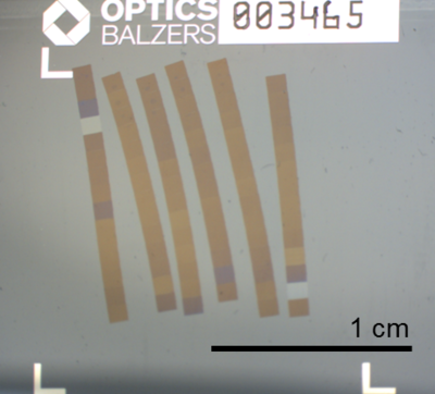
Correlative Array Tomography is an important tool for exploring the 3D structure of biological samples, new substances, etc. up to the range of several nanometers. Here, an ultra-microtome is used to cut sections of the sample and align them on a substrate (Figure 1). The sample thickness is usually a few 10 nanometers. The different sections on the substrate are imaged using different techniques (light microscope, REM, TEM). Image processing techniques are then used to segment the collected data and develop a corresponding 3D model. Manual handling of such small-sized and fragile object is basically not possible. Furthermore, structural elucidation of a complete muscle cell or an organic transistor requires several thousand sections in high-quality levels and a specific thickness.
This is why the IAI is dedicated to the automation of sample handling techniques for Correlative Array Tomography. This basically involves three major tasks: determining the section thickness, handling of the substrate, and handling of the sections.
|
|
Figure 1: Ligament sections on a glass substrate |
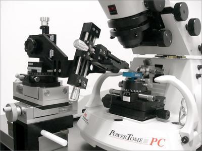
Figure 2: Manipulator for the handling of substrates on an ultra-microtome |
Digital image processing techniques are being used to determine the thickness of the sample sections. As for the substrate handling, appropriate manipulators have been developed. And fluidic manipulation methods and self-organization procedures are currently being evaluated by the IAI for the handling of the sample sections.