
CSS-CCM – Chromatic swept-source laser scanning for a confocal 3D slit lamp microscopy of the cornea
- Contact:
- Project Group:
- Funding:
DFG
- Partner:
Rostock University Medical Center
- Startdate:
2022
- Enddate:
2025
The project aims to develop a novel method for in vivo imaging of the cellular morphology of the cornea. The motivation – and expertise – for the proposed project is based on the findings obtained in the DFG project 3D-CCM, in which methods for three-dimensional in vivo confocal microscopy of the cornea with cellular resolution were developed. The project results have shown that volume data significantly increase the possibilities and standards of visual or quantitative exploration of the cellular morphology of the cornea.
The basis for 3D data acquisition in the above-mentioned project was the defined positioning of a microscope objective utilizing a piezo actuator. In combination with the frame rate of the scanner, the acquisition time for a depth scan through the entire cornea is up to 20 s. Motion artifacts in the images require complex correction algorithms for a volume reconstruction. The high operating voltage of the piezo actuator is a further drawback for a clinical application.
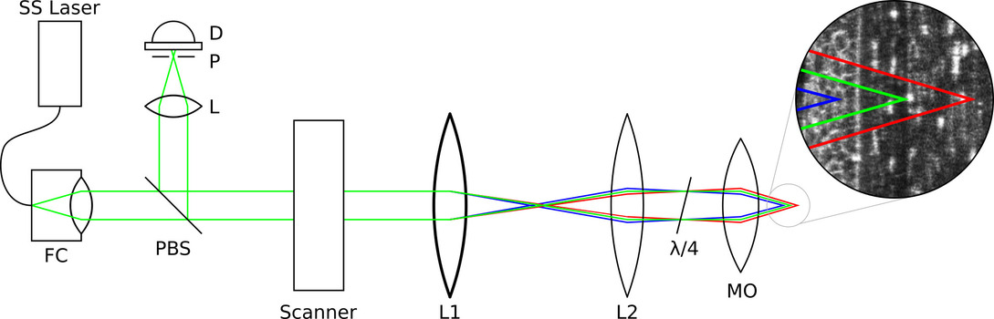
The proposed project addresses these deficits and focuses on 3D data acquisition in a completely new way, exploiting induced chromatic aberration with a swept-source laser. These narrow-band lasers periodically change their central wavelength within a large wavelength range with frequencies from a few kHz up to currently about 4 MHz.
Depending on the aberrative optics used and the oscillating wavelength of the swept-source laser, the focal depth changes in a well-defined manner. This approach entirely eliminates the need for mechanical actuators for focus positioning. It is extremely fast and, when combined with dedicated x-y scanning, allows the acquisition of a sectional volume extending through the entire corneal thickness in less than one second. For future perspective, this technology also allows the acquisition of multiple volumes per second and thus volumetric exploration of the corneal tissue using arbitrarily oriented sectional images immediately during the acquisition process. Dedicated line scanning instead of x-y scanning further improves the frame rate many times over. Thus, images can be generated without motion artifacts in image planes analogous to a slit lamp, but with cellular resolution.
This project lays the foundations for a completely novel method of high-resolution quantitative slit lamp microscopy, with great potential for improved diagnostic analyses of surface diseases of the eye. Translation of the technology into other medical fields such as dermatology for the characterization of epithelial tissue is possible, too. Due to the great and far-reaching potential of this technology, a corresponding patent specification is in preparation.
Team:
Dr. Stephan Allgeier
Karlsruhe Institute of Technology (KIT), Institute for Automation and Applied Informatics
Dr. Karsten Sperlich, Prof. Dr. Oliver Stachs
Rostock University Medical Center, Department of Ophthalmology